Microscope Imaging Facility
Our facility is located on the second floor of Taylor Science Center. Please browse our Instruments section and contact the Director if you want training or assistance.
Instruments
In 2023 we purchased a Hitachi SU3900 SEM. The system is robust and easy to use. We have many different detectors: Secondary Electrons, Backscattered Electrons, Variable Pressure, and Cathodoluminescence. Furthermore, we have 2 EDAX detectors: EDS (Octane Elite Super) and EBSD (Velocity Super). We also have support equipment to enable us to prepare and image biological samples, inorganics, metallic, geological, and others.
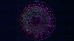
In 2024 we acquired a Nikon AXR LSCM. The system utilizes 4 lasers (405/488/561/640) to image fluorescent samples. The combination of motorized stage, resonance scanner, hybrid detectors, and user-friendly software allows for rapid and diverse imaging of samples. The system is also capable of DIC and epifluorescence imaging. Additionally, we have advanced analysis software (both Nikon and Imaris) to enable faster and more complex data collection. 4x/20x/40x LWD water/60x oil objectives.
The Nikon Ti2A is our epifluorescence microscope (also capable of brightfield). It’s easy to use, can take high resolution images or movies, and utilizes 4 filter cubes (DAPI/GFP/Texas red/Cy5). 4x/10x/20x objectives.
The Nikon AZ100 is our low magnification, high resolution brightfield microscope. This system is well suited for complex, large samples (such as plant matter or rocks). It can utilize both transmitted or reflected light, and can take high resolution images or movies. 0.5x/1x/5x objectives.
We have some additional space for sample preparation directly related to microscopy. These include a cryostat, fume hood, sputter coaters, polishing wheels, critical point dryer, and imagine analyses computers/software. The space is reserved for final preparation of samples for imaging, not for experiments.
Access and Training
All instruments are open to all faculty members and their students free of charge (does not include reagents/consumables needed to prepare your sample for microscopy). All collaborators from other institutions are welcome so long as the primary user is a Hamilton faculty member. If you need training or assistance, please contact Kyle Martin (ksmartin@hamilton.edu)